

Mammography
Early detection of breast cancer
Mammography is a special X-ray examination of the breast. It is aimed at early detection of breast cancer. Mammography is especially suited for detection of non-palpable tumours. It is possible to identify even tiny calcifications in the breast tissue. The so-called microcalcifications are often a sign of breast cancer. Small tumours can be detected starting from the size of 5 mm in diameter. At this early stage, the prospects of healing are extremely good.
As a rule, it cannot be decided immediately, whether the finding is benign or malignant. In this case, it is necessary to perform additional examinations, as e.g. sonography (ultrasound), magnetic resonance imaging (MR mammography) or biopsy. Only then it can be clearly seen, whether it is a benign or a malignant tumour.
In which cases is it necessary to perform a clarifying (curative) mammography?
- in case of a node or a hardening
- in case of unusual pains or skin changes
- nipple discharge
- lymph nodes in the armpit
- check-ups after breast cancer
- in case of strong genetic predisposition
Healthy women between 50 and 69 years of age are invited to mammography every two years in the scope of the national mammography screening programme. At this age, the risk of developing breast cancer is relatively high.
How is the examination performed?
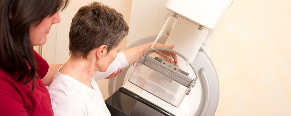
During mammography, the patient stands in front of the examination device. The medical technical radiology assistant (MTRA) discusses the whole process with the woman. She herself should define the intensity of pressure on the breast. The breast will be then cautiously compressed with a Plexiglas plate. Before the pressure gets too strong, the device stops and the recording is made. Immediately thereafter, the pressure decreases automatically.
It is necessary to know that the compression is important for a good image quality. When the breast is compressed, the radiation dose is significantly lower.
There are two images produced: a top-to-bottom view and an oblique side view. After that the images are post-processed and transferred to the report station. It takes only a few minutes. The doctor performs a tactile examination and then discusses the results with the patient. If necessary, additional examinations are carried out, e.g. mamma sonography.
Preparation for the examination
Mamography should be carried out during the first half of the menstrual cycle, about a week after the last period. During this phase of the cycle, the glandular tissue is somewhat loosened, which helps to improve the quality of the images on the one hand and makes the breast not so pressure-sensitive on the other hand.
Please do not use any creams or powders!
If you underwent a mammography in the past, bring along the previous images. Due to the fact that mammography employs X-ray radiation, the possibility of a pregnancy should be excluded.