

Magnetic resonance imaging (MRI)
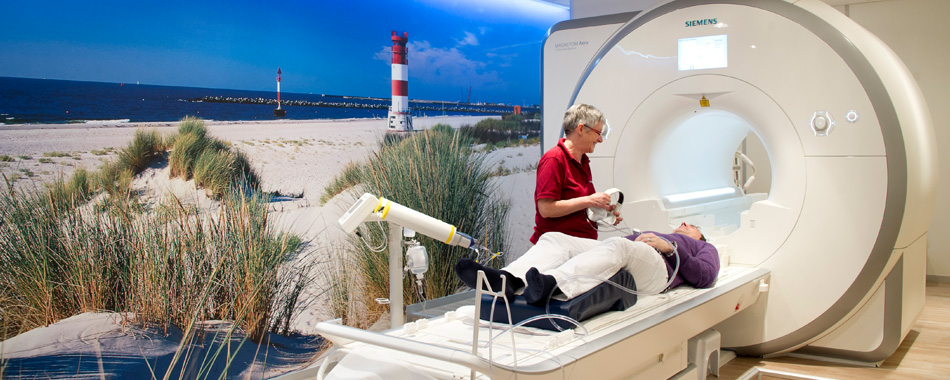
Magnetic resonance imaging does not employ any X-rays, but a strong magnetic field and radio waves instead. The heart of a magnetic resonance scanner is built up by an electromagnet weighing several tons and having a tubular opening, which the patient table will slide into. Within a short time, it is possible to make sectional images of every region of the body. Using the digital data, a computer produces images of the examined regions of the body, which will be then analysed by a radiologist.
The brain and the spinal column, the internal organs (except for the lungs) and also the muscles and joints can be most easily detected. Meanwhile, it is even possible to take images of moving organs, such as the beating heart.The further important area of application of magnetic resonance imaging is the precise depiction of blood vessels, early detection of tumours, as well as insights into the body metabolism. The virtual method of image analysis will show the referring colleagues and the patients the processes that go on inside the body.
The advantage of magnetic resonance imaging is that it is a gentle, practically risk-free method of examination. Due to low radiation exposure, even children and pregnant women can be examined. And if the patient does not tolerate iodine-containing contrast media, e.g. those applied in computed tomography, the radiologist can often choose magnetic resonance imaging.
Information on examinations of particular body parts:
Brain MRI
Brain MRI
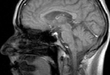
Magnetic resonance imaging (MRI) is a gentle and X-ray free examination method for producing non-overlapping and contrast-rich section images of the brain. Comfortably lying on your back, you will slide inside the examination tube after placing the head on a special device, an examination coil.
The modality of the examination is particularly suited for detecting tumours, architecture disturbances in clarification of epilepsy, vascular malformations, bleedings and circulatory disorders.
MRI is the method of choice for depicting the centres of demyelination and inflammation in the brain (MS clarification).
With the help of our modern 1,5 Tesla MRI scanner (Avanto) it is also possible to exactly distinguish between old and fresh circulatory disorders by means of special sequences (perfusion- and diffusion-weighed imaging). This information is especially important to initiate a treatment for patients at the hospital early enough.
MR angiography
MR angiography
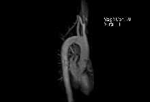
MR angiography is a gentle possibility of depicting arterial and venous vessels without X-ray exposure. In the past, catheters were inserted into arteries or veins in respective body regions with the risk of vascular injury (bleeding) or embolism (stroke). Today there is no risk at all!
Using this new technique, we are able of producing high-resolution vascular images of all regions of the body except for coronary vessels. With the help of our highly modern 1,5 Tesla Avanto scanner, we are even able to depict aortas and leg and foot vessels during examination procedure.
For that purpose, the patient will be placed between the specially developed coils and then put inside the scanner. A gadolinium-based contrast agent will then be injected through the previously fixed puncture cannulae, in order to make the vessels of the body visible.
For examination of head vessels it is necessary to administer a contrast agent.
With the help of this examination, it is possible to depict narrowed (stenosis) or dilated (aneurysms) vessels, blood clots (thrombi) or vascular occlusions.
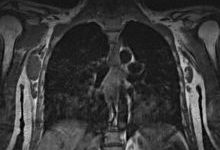
Neck MRI
Neck MRI
Magnetic resonance imaging (MRI) is a gentle and X-ray free examination method for producing non-overlapping and contrast-rich section images of the neck organs. For that purpose, the patient will lie on his back. An examination coil will be placed onto the front soft tissues of the neck, and the patient will then slide into the scanner.
With the help of this method it is possible to clearly evaluate lymph nodes, the tongue, the larynx and the base of the tongue and to exclude inflammatory or tumorous changes.
Thorax MRI
Thorax MRI
Magnetic resonance imaging (MRI) is a gentle and X-ray free examination method for producing non-overlapping and contrast-rich section images of the thoracic wall, axilla and mediastinum. For that purpose, the patient will lie on his back. An examination coil will be placed onto the front wall, and the patient will then slide into the scanner.
By means of this examination, it is possible to detect inflammations, tumours and metastases of the spinal column, thoracic wall and axillas. However, a detailed evaluation of the lung parenchyma cannot be done with the help of MRI, even though our modern 1,5 Tesla Avanto scanner can depict lung metastases from the size of 10 mm. The search of lung metastases should be carried out by means of computed tomography (CT).
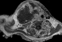
Abdominal MRI
Abdominal MRI
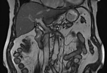
Magnetic resonance imaging (MRI) is a gentle possibility of depicting all abdominal organs vessels without X-ray exposure. With this examination modality, high-resolution and non-overlapping images of the internal organs are produced.
For that purpose, the patient will be comfortably placed on his back, and a special system of coils will be arranged in the examination area, in order to obtain qualitative images. Depending on the examination protocol, the patient can breathe during the examination, or hold his breath several times for 10-20 seconds, after the respective pause. If necessary, at the end of the examination a gadolinium-based contrast agent will be administered.
Especially the liver MRI is now preferred to computer tomography (CT) for the search of metastases and tumour aftercare, due to the fact that a lot of liver-specific contrast agents have been introduced in recent years.
With the help of these contrast agents, as well as specific dynamic examination protocols of our modern 1,5 Tesla Scanner, today we are able to distinguish between benign and malignant changes. This method allows the best possible clarification not only of the liver, but also of the kidneys, adrenal glands, pancreas, spleen and sex organs.
In addition, in out practice we can carry out high-resolution examinations of the small intestine (MR-Sellink), in order to be able to detect inflammations (Crohn's disease), ingrowths, passage obstructions or intra-abdominal cyst formation (e.g. endometriosis).
This examination is very time-consuming, because before the beginning of the examination the patients have to drink a special mannitol solution in small sips, spread over a period of 40 minutes.
We also have new sequences at our disposal, which allow us to produce high-resolution images of the bile and pancreatic ducts (MRCP).
Spine/joint MRI
Spine/joint MRI
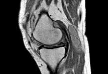
Magnetic resonance imaging (MRI) of the spine and joints produces non-overlapping, excellent images without the use of X-rays.
This is the only method depicting cartilage and ligamentous structures and also intervertebral discs and spinal cord structures in adequate quality and resolution.
For that purpose, the patient will be comfortably placed either on his back, or on his belly, as far as possible. The respective extremity (joint) will be wrapped into a special examination coil. And finally, the patient will be placed into the scanner.